

Here is a look at the current screen:

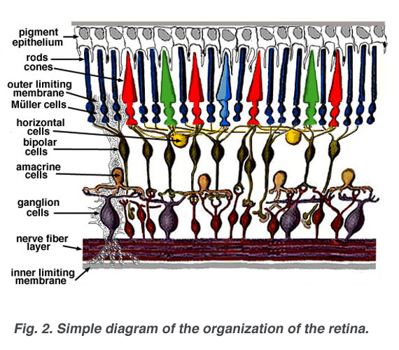
Colored circles are Red, Green and Blue Cone photoreceptors shown below at the outer layer of retina.
From: http://webvision.med.utah.edu/Wong.html
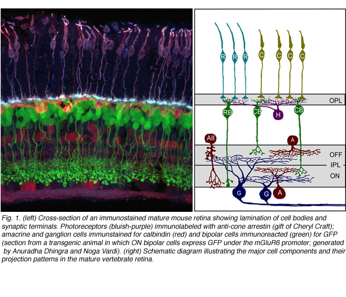
The above layers organize into distinct laminae summed up below.
1) In the outer retina, photoreceptors contact horizontal cells and bipolar cells within the outer plexiform layer (OPL).
2) Within the inner retina, synapses between retinal ganglion cells and their presynaptic partners, the amacrine and bipolar interneurons are localized to the inner plexiform layer (IPL).
Connections that depolarized upon increase in illumination (ON) occupy approximately the inner half of the IPL,
whereas connectivity of cells hyperpolarized (OFF) by light is confined to the outer half of the IPL.
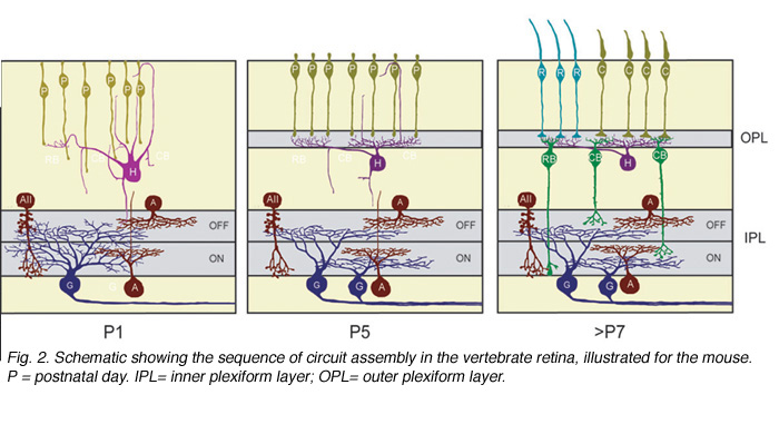
Above is shown the three major phases of wiring.
1) In the IPL, retinal ganglion cells and amacrine cells form the earliest functional circuits.
2) Forming the OPL, horizontal cells and photoreceptors connect.
3) Bipolar cells are born, interconnect vertical networks with ganglion cells are established.
In more detail is the following early development showing the "Nerve fiber layer" at the bottom of drawing:
From: http://webvision.med.utah.edu/anatomy.html
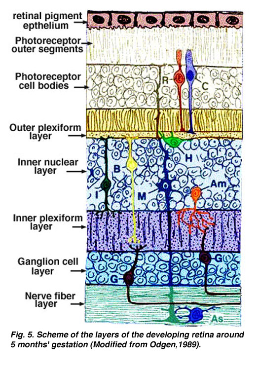
The retina develops in an inside to outside manner: ganglion cells are formed first and photoreceptors cells become fully mature last. The Vision System program can be born with cells each with a small Self-Learning memory where confidence increases when it behaves as would a real cell.
-